Bone Cross Section : Cross Section Neck Through Hyoid Bone Royalty Free Vector - The head of the cutting cone consists of osteoclasts that resorb the bone.
Bone Cross Section : Cross Section Neck Through Hyoid Bone Royalty Free Vector - The head of the cutting cone consists of osteoclasts that resorb the bone.. If you know the orientation of the section, you can easily identify the bones because the pubic bone sits anteriorly in the pelvis. This photo shows a cross section through bone. Erythrocytes, or red blood cells, are by far the predominant cell type in the blood smear. Cancellous bone, light, porous bone enclosing numerous large spaces that give a honeycombed or spongy appearance. Can you identify the primary and secondary haversian systems, central canals and bone lamellae?
Cancellous bone, light, porous bone enclosing numerous large spaces that give a honeycombed or spongy appearance. This photo shows a cross section through bone. Each epiphysis meets the diaphysis at the metaphysis, the narrow area that contains the epiphyseal plate (growth plate), a layer of hyaline (transparent) cartilage in a growing. They form the acetabulum, which is represented by the reddish semilunar shape. Red marrow fills the spaces in the spongy bone.
Cross Section Of A Femur Bone Showing The Anatomical Structure Including Cancellous Bone And Marrow Stock Photo Picture And Rights Managed Image Pic Sjb 4378 2939 Agefotostock from previews.agefotostock.com The wider section at each end of the bone is called the epiphysis (plural = epiphyses), which is filled with spongy bone. Cuttings cones, or sheets of osteoclasts, bore holes through the hard bone, leaving tunnels, which appear in cross section as cavities. The head of the cutting cone consists of osteoclasts that resorb the bone. If you know the orientation of the section, you can easily identify the bones because the pubic bone sits anteriorly in the pelvis. This photo shows a cross section through bone. Can you identify the primary and secondary haversian systems, central canals and bone lamellae? They form the acetabulum, which is represented by the reddish semilunar shape. Red marrow fills the spaces in the spongy bone.
If you know the orientation of the section, you can easily identify the bones because the pubic bone sits anteriorly in the pelvis.
They form the acetabulum, which is represented by the reddish semilunar shape. Cortical bone remodeling proceeds via cutting cones and is similar to processes in other hard biological tissues. Can you identify the primary and secondary haversian systems, central canals and bone lamellae? The scaphoid bone is the largest bone of the proximal row of wrist bones, its long axis being from above downward, lateralward, and forward. The next section will discuss the identification of the immature cells of the bone marrow. If you know the orientation of the section, you can easily identify the bones because the pubic bone sits anteriorly in the pelvis. Erythrocytes, or red blood cells, are by far the predominant cell type in the blood smear. This is a high power photo of a single haversian system. The wider section at each end of the bone is called the epiphysis (plural = epiphyses), which is filled with spongy bone. The bone is shaped like the numeral 7, 1 which gives these cuts their name. Cancellous bone, light, porous bone enclosing numerous large spaces that give a honeycombed or spongy appearance. Red marrow fills the spaces in the spongy bone. Cuttings cones, or sheets of osteoclasts, bore holes through the hard bone, leaving tunnels, which appear in cross section as cavities.
Red marrow fills the spaces in the spongy bone. Erythrocytes, or red blood cells, are by far the predominant cell type in the blood smear. This is a high power photo of a single haversian system. The scaphoid bone is the largest bone of the proximal row of wrist bones, its long axis being from above downward, lateralward, and forward. Cortical bone remodeling proceeds via cutting cones and is similar to processes in other hard biological tissues.
Cross Section Neck Through Hyoid Bone Royalty Free Vector from cdn1.vectorstock.com They form the acetabulum, which is represented by the reddish semilunar shape. Red marrow fills the spaces in the spongy bone. Online medical dictionary and glossary with medical definitions, s listing. Can you identify the primary and secondary haversian systems, central canals and bone lamellae? If you know the orientation of the section, you can easily identify the bones because the pubic bone sits anteriorly in the pelvis. The next section will discuss the identification of the immature cells of the bone marrow. Erythrocytes, or red blood cells, are by far the predominant cell type in the blood smear. The head of the cutting cone consists of osteoclasts that resorb the bone.
Red marrow fills the spaces in the spongy bone.
This photo shows a cross section through bone. The wider section at each end of the bone is called the epiphysis (plural = epiphyses), which is filled with spongy bone. Cancellous bone, light, porous bone enclosing numerous large spaces that give a honeycombed or spongy appearance. Each epiphysis meets the diaphysis at the metaphysis, the narrow area that contains the epiphyseal plate (growth plate), a layer of hyaline (transparent) cartilage in a growing. Red marrow fills the spaces in the spongy bone. Cortical bone remodeling proceeds via cutting cones and is similar to processes in other hard biological tissues. This is a high power photo of a single haversian system. The head of the cutting cone consists of osteoclasts that resorb the bone. If you know the orientation of the section, you can easily identify the bones because the pubic bone sits anteriorly in the pelvis. Erythrocytes, or red blood cells, are by far the predominant cell type in the blood smear. The bone is shaped like the numeral 7, 1 which gives these cuts their name. The next section will discuss the identification of the immature cells of the bone marrow. Can you identify the primary and secondary haversian systems, central canals and bone lamellae?
The head of the cutting cone consists of osteoclasts that resorb the bone. Online medical dictionary and glossary with medical definitions, s listing. The wider section at each end of the bone is called the epiphysis (plural = epiphyses), which is filled with spongy bone. The bone is shaped like the numeral 7, 1 which gives these cuts their name. This is a high power photo of a single haversian system.
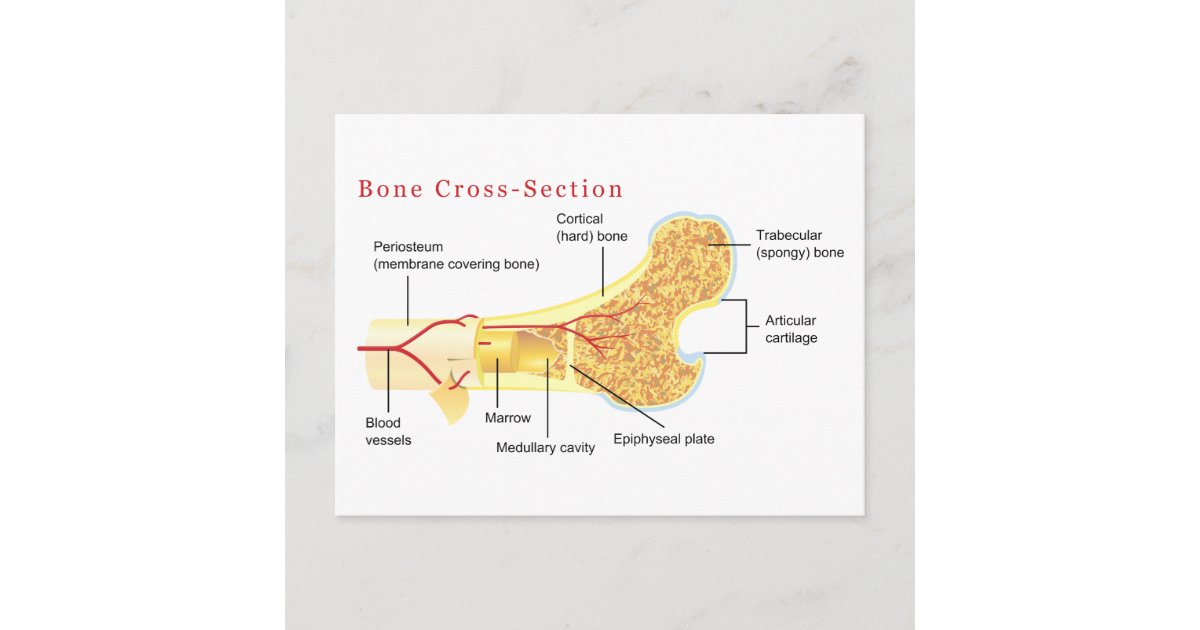
Bone Cross Section Diagram Postcard Zazzle Com from rlv.zcache.com This photo shows a cross section through bone. Cancellous bone, light, porous bone enclosing numerous large spaces that give a honeycombed or spongy appearance. They form the acetabulum, which is represented by the reddish semilunar shape. The next section will discuss the identification of the immature cells of the bone marrow. Red marrow fills the spaces in the spongy bone. Can you identify the primary and secondary haversian systems, central canals and bone lamellae? Online medical dictionary and glossary with medical definitions, s listing. The bone is shaped like the numeral 7, 1 which gives these cuts their name.
The head of the cutting cone consists of osteoclasts that resorb the bone.
Erythrocytes, or red blood cells, are by far the predominant cell type in the blood smear. This photo shows a cross section through bone. The next section will discuss the identification of the immature cells of the bone marrow. Cortical bone remodeling proceeds via cutting cones and is similar to processes in other hard biological tissues. Cancellous bone, light, porous bone enclosing numerous large spaces that give a honeycombed or spongy appearance. If you know the orientation of the section, you can easily identify the bones because the pubic bone sits anteriorly in the pelvis. The wider section at each end of the bone is called the epiphysis (plural = epiphyses), which is filled with spongy bone. The head of the cutting cone consists of osteoclasts that resorb the bone. Can you identify the primary and secondary haversian systems, central canals and bone lamellae? Red marrow fills the spaces in the spongy bone. This is a high power photo of a single haversian system. Cuttings cones, or sheets of osteoclasts, bore holes through the hard bone, leaving tunnels, which appear in cross section as cavities. The bone is shaped like the numeral 7, 1 which gives these cuts their name.


Comments
Post a Comment